Alzheimer’s disease can develop for years before a person notices the first memory problems. But is it possible to detect signs of future cognitive decline while the brain is still managing everyday tasks? Scientists at Stanford Medicine found that certain patterns of brain activity may be associated with the risk of declining memory and thinking long before noticeable symptoms appear.
Researchers compared functional brain connectivity in people with Alzheimer’s disease and older adults with exceptionally good memory. They then studied brain activity in adults without cognitive impairment and found that the more its patterns resembled the profile of people with Alzheimer’s disease, the faster their cognitive abilities declined over the following years.
The findings, published on November 4, 2026, in Science Advances, open up new possibilities for early assessment of cognitive decline risk. However, as Stanford Medicine emphasizes, the proposed measure is still a research tool. It cannot be used to make a diagnosis or reliably predict whether a particular person will develop Alzheimer’s disease.
Why compare people with Alzheimer’s and superagers
Age alone does not explain why some people retain excellent memory while others experience a marked decline. The researchers selected two groups representing different forms of cognitive aging: 180 superagers and 110 people with Alzheimer’s disease. All participants were at least 75 years old.
In this study, superagers were defined as people whose memory was comparable to the level typical of 45-year-olds. Their executive functions, including the ability to plan actions and manage attention, remained within the normal range for their age. This definition does not mean that all of a person’s abilities are equivalent to those of someone of average age.
The comparison made it possible to identify a characteristic profile of functional connectivity. Based on it, the authors created the ARC measure, whose name can be translated as “Alzheimer’s Resilience and Vulnerability Connectome.” Here, connectome means a map of connections between brain regions.
The idea behind this approach is to use information from both ends of the spectrum: taking into account brain patterns associated with the disease and those associated with unusually well-preserved memory in older age.
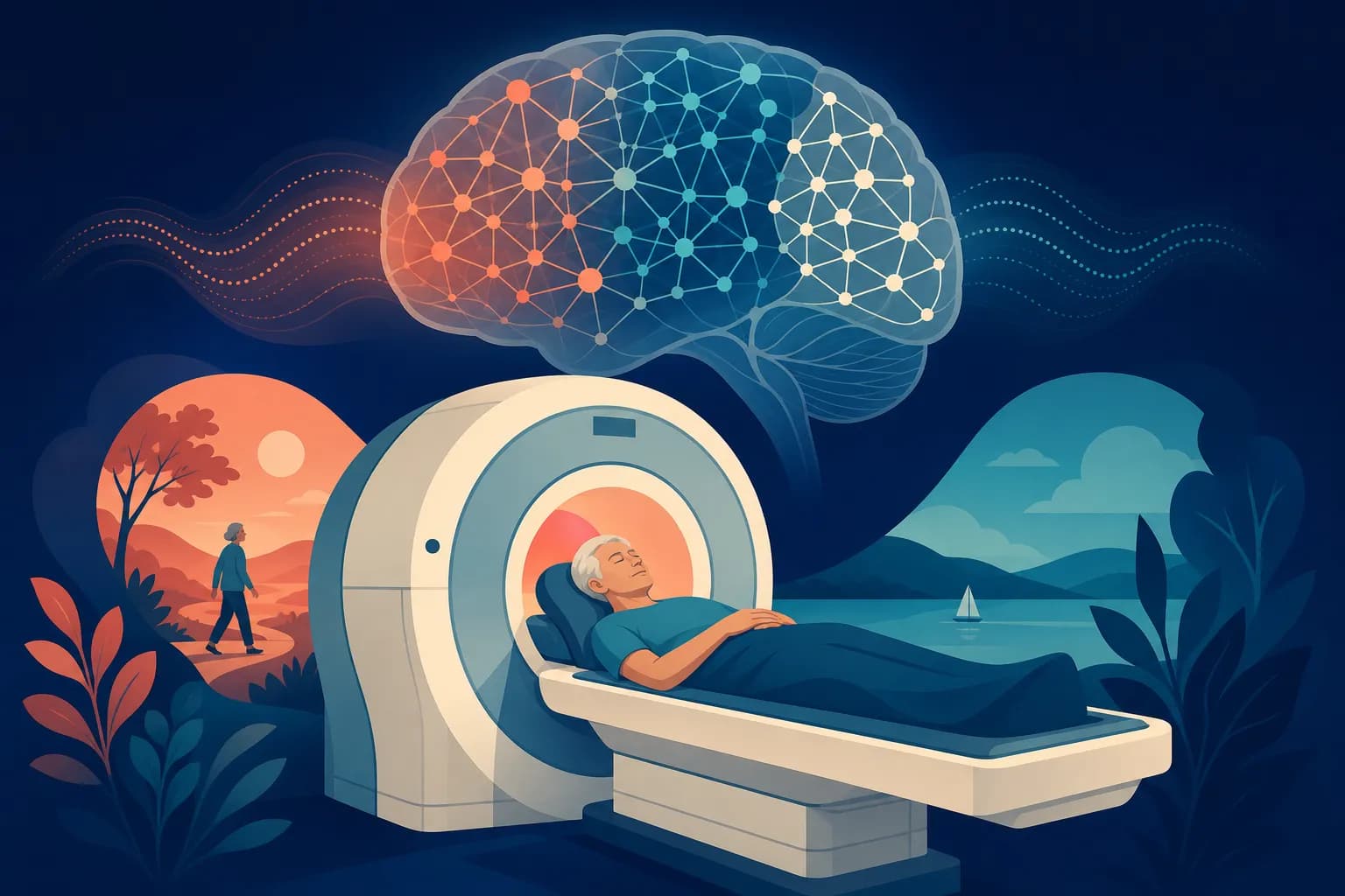
What functional brain MRI measures
The study used functional MRI scans taken at rest. During this type of scan, a person does not perform a specific memory task. The method records changes in the signal associated with blood oxygen levels and uses them to indirectly assess brain activity.
When signals in different regions change in a coordinated way, researchers refer to this as functional connectivity. This is a statistical coordination of activity; it does not necessarily indicate a direct anatomical connection between two areas of the brain.
According to the study abstract, the identified profile involved prefrontal, temporal, and insular networks. These regions take part in a range of processes, including memory, cognitive control, and the processing of relevant information. ARC describes a distributed pattern of interaction that cannot be reduced to the activity of a single region.
This explains the interest in functional MRI for early risk detection: it makes it possible to study how coordinated brain systems are, even when cognitive test results are still normal.
Even before memory problems appear, interactions between different areas of the brain in some people begin to resemble the pattern characteristic of Alzheimer’s disease.
The more pronounced this similarity, the higher the risk of subsequent cognitive decline.
What the researchers found in people without memory impairment
After developing the model, the authors turned to the main question: is this profile associated with future changes in people who do not yet have cognitive impairment? The analysis included a primary sample of 818 people and an independent validation sample of 497 people.
Participants with profiles resembling those of superagers and people with Alzheimer’s disease had comparable baseline cognitive scores. But their condition changed differently over time.
Similarity to the disease profile was associated with faster cognitive decline, greater accumulation of beta-amyloid, and an increased risk of subsequently developing Alzheimer’s disease.
Beta-amyloid is a protein fragment that can accumulate in the brain and form plaques characteristic of the disease. The link between ARC and its accumulation adds to the findings on memory: the measure was also associated with a biological process related to the disease.
The scientific value of the findings lies in the difference between subsequent trajectories despite similar starting test scores. At the same time, the study does not establish that this connectivity pattern causes the disease. It may reflect early changes or other aspects of brain vulnerability.
What the follow-up period of up to 12 years means
Stanford’s report says that participants were followed for up to 12 years, although most of the data covered five to six years. This is the maximum follow-up period stated in the university’s report, not a uniform forecast period for every person.
The AAAS press release gives a different range: five to 15 years. Therefore, the phrase “12 years ahead” should be understood with the source and the varying follow-up periods in mind.
The practical significance of the findings remains: the baseline brain connectivity profile was associated with events that occurred years later. However, this does not mean that a single scan can reveal whether someone will develop the disease by a particular date.
How this discovery could help with early Alzheimer’s diagnosis
The most immediate potential use of ARC is in prevention research. If the measure helps identify a group of cognitively healthy people who are more likely to experience decline, it could be studied as a tool for selecting participants for clinical trials. For now, this remains a possibility that needs further testing.
Before it can be used in routine practice, researchers will need to understand how much ARC improves prediction compared with available clinical information and other biomarkers. They will also need to establish which medical decisions would actually benefit from the results of such an assessment.
Searching for risk markers and testing prevention methods are different tasks. This distinction helps put both findings about aspirin and dementia and comprehensive approaches such as the Bredesen Protocol into perspective: an association with a future outcome does not, by itself, show which intervention can change that outcome.
If memory is noticeably worsening and interfering with everyday life, it makes more sense to seek a clinical assessment than to look for an experimental measure. For someone without impairment, this study does not yet provide a reason to undergo functional MRI for predictive purposes. The value of an early signal will depend on whether a proven way to use it to improve outcomes becomes available.
What does an AUC of 0.85 mean in this study?
It describes the model’s ability to distinguish superagers from people with an established Alzheimer’s diagnosis in the original sample. Put simply, in about 85% of randomly selected pairs from these two groups, the model correctly ranked participants by how closely they resembled the disease profile. This does not mean an 85% accuracy rate for predicting Alzheimer’s in a healthy person 12 years in advance.
Can ARC be improved through memory training or brain stimulation?
It is not yet known whether this measure can be reliably changed or whether changing it would be beneficial. The possibility of influencing it remains a hypothesis. This study did not demonstrate that training or brain stimulation can prevent cognitive decline.
Do Alzheimer’s disease and dementia mean the same thing?
No. Dementia refers to a syndrome of significant cognitive decline that interferes with independent everyday life. Alzheimer’s disease is one of its causes. Therefore, findings from a study focused on Alzheimer’s cannot automatically be applied to all forms of dementia.